Affected pathways in Duchenne muscular dystrophy (WP5356)
Homo sapiens

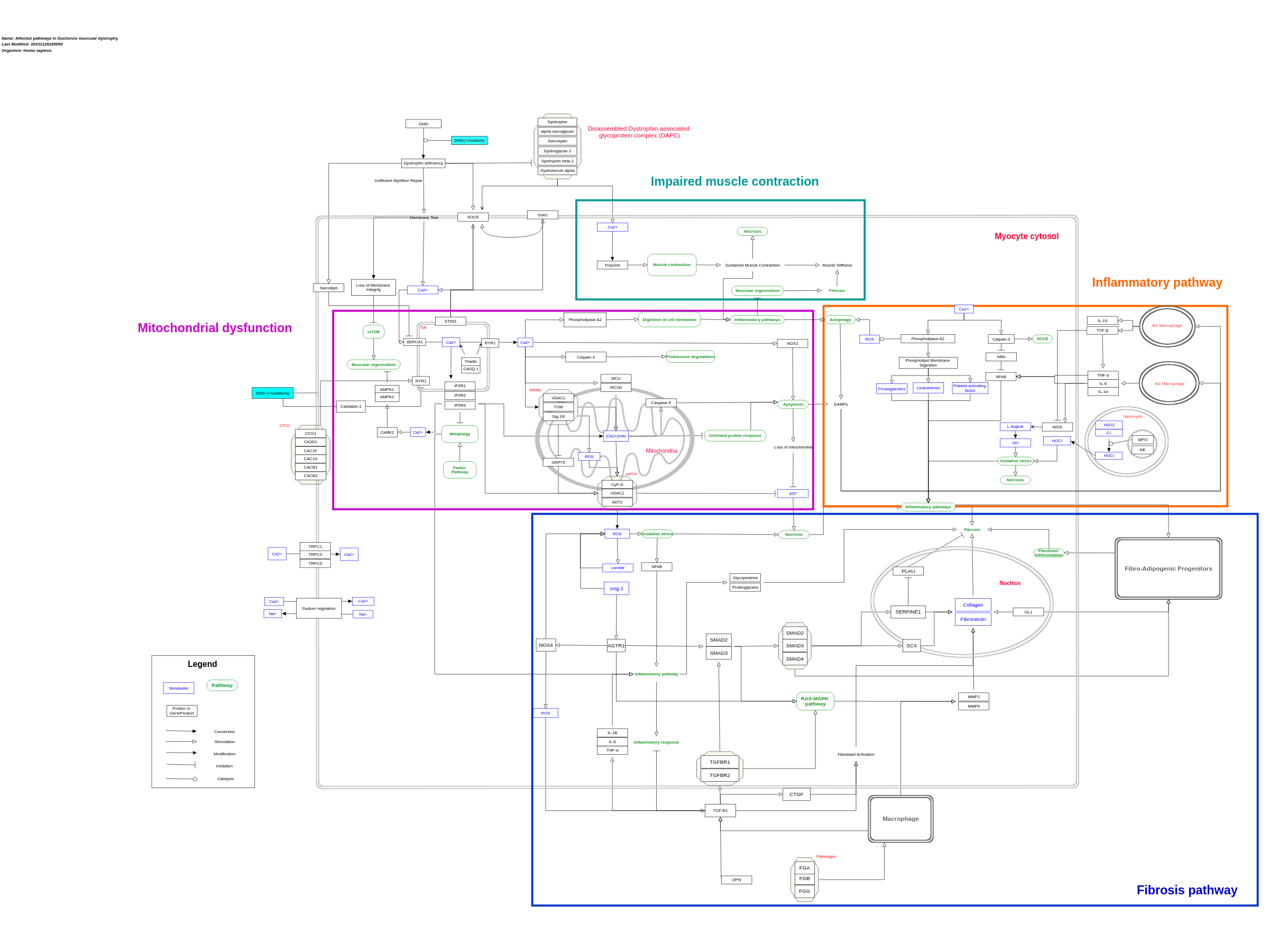
DMD (Duchenne Muscular Dystrophy) is a genetic disorder that primarily affects muscles in the body, causing progressive muscle weakness and wasting. It is caused by mutations in the DMD gene, which results in a deficiency or absence of the protein dystrophin, leading to muscle degeneration. DMD is characterized by abnormal calcium levels resulting from dysfunction in the muscle cell membrane. This leads to the uncontrolled opening of the mitochondrial permeability transition pore (mPTP) which inhibits ATP synthesis and thus, drives the cell into apoptosis. This influx activates a cascade of harmful events, including increased production of reactive oxygen species (ROS) and activation of proteases that can damage the cell structure of muscle fibers. The lack of the dystrophin protein affects essential components for muscle contraction namely the Disassembled Dystrophin-associated glycoprotein complex (DAPC) which disturbs the normal contraction-relaxation process of the muscle in DMD. Sustained contractions occur due to the high calcium influx causing muscle stiffness and fibrosis, which are known characteristics of DMD. Indeed, fibrosis is commonly stimulated in dystrophic muscle cells as a result of the upregulation of several pro-fibrotic transcription factors such as SERPINE1, SCX and GL1. Hence, excessive amounts of collagen and fibronectin are produced, enhancing fibrosis. All these events cause chronic inflammation in the muscle cell, attracting pro-inflammatory cytokines, chemokines and other inflammatory mediators. The chronic inflammation in DMD can further perpetuate muscle degeneration, fibrosis, and impaired muscle function. Acknowledgments: Bria Jackson, Amaia Alvarez van Schie, Otto Rämö, Tuneille Adelaar
For a description of pathway objects, see the WikiPathways Legend.
Authors
Pauladewenter , Aishwarya Iyer , Egon Willighagen , Alex Pico , Kristina Hanspers , Lars Willighagen , Eric Weitz , Tooba Abbassi-Daloii , and Daniela DiglesActivity

Discuss this pathway
Check for ongoing discussions or start your own.
Cited In
Are you planning to include this pathway in your next publication? See How to Cite and add a link here to your paper once it's online.
Organisms
Homo sapiensCommunities
Diseases Rare DiseasesAnnotations
Cell Type Ontology
muscle cell macrophage inflammatory macrophage mesenchymal stem cell alternatively activated macrophageDisease Ontology
muscular disease Duchenne muscular dystrophyPathway Ontology
muscular disease pathway altered calcium/calcium-mediated signaling pathway disease pathway| Label | Type | Compact URI | Comment |
|---|---|---|---|
| Lactate | Metabolite | chebi:24996 | |
| [Ca2+]mito | Metabolite | hmdb:HMDB0000464 | |
| Ca2+ | Metabolite | hmdb:HMDB0000464 | |
| ROS | Metabolite | wikidata:Q424361 | |
| ATP | Metabolite | chebi:30616 | |
| Fibronectin | Metabolite | chebi:5058 | |
| Collagen | Metabolite | chebi:3815 | |
| Ang 2 | Metabolite | pubchem.compound:172198 | 'angiotensin 2 (Ang 2)' Peptide hormone; 'Asp-Arg-Val-Tyr-Ile-His-Pro-Phe Angiotensin I is converted to angiotensin II (AII) through removal of two C-terminal residues by the enzyme angiotensin-converting enzyme (ACE), primarily through ACE within the lung (but also present in endothelial cells, kidney epithelial cells, and the brain). Angiotensin II acts on the central nervous system to increase vasopressin production, and also acts on venous and arterial smooth muscle to cause vasoconstriction. Angiotensin II also increases aldosterone secretion; it therefore acts as an endocrine, autocrine/paracrine, and intracrine hormone.' Source: 'https://en.wikipedia.org/wiki/Angiotensin#Angiotensin_II' |
| Platelet-activating factor | Metabolite | hmdb:HMDB0062195 | |
| Na+ | Metabolite | hmdb:HMDB0000588 | |
| Cl- | Metabolite | hmdb:HMDB0000492 | |
| NO | Metabolite | hmdb:HMDB0003378 | |
| H2O2 | Metabolite | hmdb:HMDB0003125 | |
| HOCl | Metabolite | hmdb:HMDB0001050 | Hypochlorous acid (HOCl) is a type of reactive oxygen species which is produced within neutrophils, and can enter the muscle tissue where it promotes oxidative stress |
| L-Arginine | Metabolite | hmdb:HMDB0000517 | |
| HOCl | Metabolite | hmdb:HMDB0001050 | |
| RYR1 | GeneProduct | hgnc.symbol:RYR1 | |
| NOX2 | GeneProduct | uniprot:P04839 | |
| SOCE | GeneProduct | hgnc.symbol:SARAF | |
| Dystrophin deficiency | GeneProduct | hgnc.symbol:DMD | |
| DMD | GeneProduct | hgnc.symbol:DMD | |
| DMD (+ mutation) | GeneProduct | hgnc.symbol:DMD | |
| TGFB1 | GeneProduct | ensembl:ENSG00000105329 | |
| ADT2 | GeneProduct | hgnc.symbol:SLC25A5 | |
| OPN | GeneProduct | hgnc.symbol:SPP1 | |
| TGFBR2 | GeneProduct | hgnc.symbol:TGFBR2 | |
| TGFBR1 | GeneProduct | hgnc.symbol:TGFBR1 | |
| SERPINE1 | GeneProduct | hgnc.symbol:SERPINE1 | Also known as plasminogen activator inhibitor-1 PAI-1 |
| PLAU | GeneProduct | hgnc.symbol:PLAU | Also known as Urokinase-type plasminogen inhibitor (uPA) |
| IL6 | GeneProduct | ensembl:ENSG00000136244 | In DMD, IL-6 is upregulated due to recurrent activation of the M1 macrophages by DAMPs. When upregulated for prolonged periods of time, the IL-6 will cause chronic inflammation and also reduce the population of the satellite cells that are needed for muscle regeneration. |
| IL1B | GeneProduct | ensembl:ENSG00000125538 | |
| SMAD2 | GeneProduct | hgnc.symbol:SMAD2 | |
| SMAD3 | GeneProduct | hgnc.symbol:SMAD3 | |
| NOX4 | GeneProduct | hgnc.symbol:NOX4 | |
| CTGF | GeneProduct | hgnc.symbol:CCN2 | |
| TNF | GeneProduct | ensembl:ENSG00000232810 | |
| AGTR1 | GeneProduct | hgnc.symbol:AGTR1 | |
| DTNA | GeneProduct | hgnc.symbol:DTNA | 'Dystrobrevin alpha' originally |
| SNTB1 | GeneProduct | hgnc.symbol:SNTB1 | 'Syntrophin beta-1' originally |
| SGCA | GeneProduct | hgnc.symbol:SGCA | 'alpha sarcoglycan' originally |
| SSPN | GeneProduct | hgnc.symbol:SSPN | 'Sarcospan' originally |
| DMD | GeneProduct | hgnc.symbol:DMD | 'Dystrophin' originally |
| DAG1 | GeneProduct | hgnc.symbol:DAG1 | 'Dystroglycan 1' originally |
| STIM1 | GeneProduct | ensembl:ENSG00000167323 | |
| ORAI1 | GeneProduct | ensembl:ENSG00000276045 | |
| CACNA1F | GeneProduct | ensembl:ENSG00000102001 | Voltage-dependent L-type calcium channel subunit alpha-1F |
| CACNA1S | GeneProduct | ensembl:ENSG00000081248 | Voltage-dependent L-type calcium channel subunit alpha-1S |
| CACNB2 | GeneProduct | ensembl:ENSG00000165995 | Voltage-dependent L-type calcium channel subunit beta-2 |
| CACNB1 | GeneProduct | ensembl:ENSG00000067191 | Voltage-dependent L-type calcium channel subunit beta-1. Only isoform 2 is present in skeletal muscles |
| CACNA2D1 | GeneProduct | ensembl:ENSG00000153956 | Voltage-dependent calcium channel subunit alpha-2/delta-1. Only isoform 1 is found in the skeletal muscles |
| CACNG1 | GeneProduct | ensembl:ENSG00000108878 | Voltage-dependent calcium channel subunit gamma-1 |
| DMD (+mutations) | GeneProduct | ensembl:ENSG00000198947 | |
| FKBP1A | GeneProduct | ensembl:ENSG00000088832 | 'Calstabin-1' originally. Often, there is Ca2+ leakage from the RyRs in the SR, but this process is limited by calstabin-1. Calstabin-1 is a protein which has a high affinity for RyR, stimulated by the dystrophin. However, due to DMD mutations and thus reduction of dystrophin, the Calstabin-1 no longer binds with such a high affinity to the RyR, thus not blocking the calcium leakage |
| IL1A | GeneProduct | ensembl:ENSG00000115008 | |
| IL10 | GeneProduct | ensembl:ENSG00000136634 | |
| NFkB | GeneProduct | ensembl:ENSG00000109320 | |
| NFKBIA | GeneProduct | ensembl:ENSG00000100906 | 'IκBα' in original source |
| CAPN3 | GeneProduct | ensembl:ENSG00000092529 | 'Calpain-3' in original source |
| PLA2G4A | GeneProduct | ensembl:ENSG00000116711 | 'Phospholipase A2' in original source |
| TRPC3 | GeneProduct | ensembl:ENSG00000138741 | Short transient receptor potential channel 3 |
| TRPC1 | GeneProduct | ensembl:ENSG00000144935 | Short transient receptor potential channel 1 |
| TRPC6 | GeneProduct | ensembl:ENSG00000137672 | Short transient receptor potential channel 6 |
| CASQ 1 | GeneProduct | hgnc.symbol:CASQ1 | |
| TRDN | GeneProduct | hgnc.symbol:TRDN | 'Triadin' originally |
| AMPK1 | GeneProduct | ensembl:ENSG00000132356 | |
| NE | GeneProduct | ensembl:ENSG00000197561 | Neutrophil elastase (NE) is a type of serine protease which promotes the formation of neutrophil extracellular traps (NETs), which themselves function to trap invading microbes but also cuase tissue damage as collateral. Although possibly playing a role in the pathophysiology of DMD, it is not yet confirmed if NETs contribute to the muscle damage |
| MPO | GeneProduct | ensembl:ENSG00000005381 | Myeloperoxidase (MPO) is a potent enzyme which catalyses the production of hypochlorous acid (HOCl) when hydrogen peroxide (H2O2) and chloride ions (Cl-) are present within the neutrophil. |
| FGG | GeneProduct | hgnc.symbol:FGG | |
| FGB | GeneProduct | hgnc.symbol:FGB | |
| FGA | GeneProduct | hgnc.symbol:FGA | |
| CASP9 | Protein | hgnc.symbol:CASP9 | 'Caspase 9' originally |
| VDAC1 | Protein | hgnc.symbol:VDAC1 | |
| PPIF | Protein | hgnc.symbol:PPIF | 'CyP-D' originally |
| IP3R2 | Protein | hgnc.symbol:ITPR2 | |
| IP3R3 | Protein | hgnc.symbol:ITPR3 | |
| PLA2G2A | Protein | hgnc.symbol:PLA2G2A | 'Phospholipase A2' originally |
| MMP2 | Protein | hgnc.symbol:MMP2 | |
| TOMM20 | Protein | hgnc.symbol:TOMM20 | 'TOM' originally |
| GLI1 | Protein | hgnc.symbol:GLI1 | 'GL1' originally |
| MCU | Protein | hgnc.symbol:MCU | |
| IP3R1 | Protein | uniprot:Q14643 | |
| Proteoglycans | Protein | hgnc.symbol:PRG3 | |
| SERCA1 | Protein | hgnc.symbol:ATP2A1 | |
| Glycoproteins | Protein | hgnc.symbol:GP2 | |
| TNNI1 | Protein | hgnc.symbol:TNNI1 | 'Troponin' originally |
| CAPN3 | Protein | hgnc.symbol:CAPN3 | 'Calpain-3' originally |
| MCUB | Protein | hgnc.symbol:MCUB | |
| MMP9 | Protein | hgnc.symbol:MMP9 | |
| SIGMAR1 | Protein | hgnc.symbol:SIGMAR1 | 'Sig-1R' originally |
| GRP75 | Protein | uniprot:P38646 | |
| SMAD4 | Protein | hgnc.symbol:SMAD4 | |
| SMAD2 | Protein | hgnc.symbol:SMAD2 | |
| SMAD3 | Protein | hgnc.symbol:SMAD3 | |
| SCX | Protein | hgnc.symbol:SCX | |
| SLN | Protein | ensembl:ENSG00000170290 | 'Sarcolipin' originally |
| CAMK2D | Protein | ensembl:ENSG00000145349 | 'CaMK2' originally |
| AMPK2 | Protein | ensembl:ENSG00000162409 | |
| NOS2 | Protein | ensembl:ENSG00000007171 | 'iNOS' in original source |
References
- Phospholipase A2 activity in dystrophinopathies. Lindahl M, Bäckman E, Henriksson KG, Gorospe JR, Hoffman EP. Neuromuscul Disord. 1995 May;5(3):193–9. PubMed Europe PMC Scholia
- Function and genetics of dystrophin and dystrophin-related proteins in muscle. Blake DJ, Weir A, Newey SE, Davies KE. Physiol Rev. 2002 Apr;82(2):291–329. PubMed Europe PMC Scholia
- Muscular dystrophies: genes to pathogenesis. Dalkilic I, Kunkel LM. Curr Opin Genet Dev. 2003 Jun;13(3):231–8. PubMed Europe PMC Scholia
- Lactate and oxygen constitute a fundamental regulatory mechanism in wound healing. Trabold O, Wagner S, Wicke C, Scheuenstuhl H, Hussain MZ, Rosen N, et al. Wound Repair Regen. 2003;11(6):504–9. PubMed Europe PMC Scholia
- Transcriptional and posttranscriptional regulation of the plasminogen activator system. Nagamine Y, Medcalf RL, Muñoz-Cánoves P. Thromb Haemost. 2005 Apr;93(4):661–75. PubMed Europe PMC Scholia
- New roles of calsequestrin and triadin in cardiac muscle. Knollmann BC. J Physiol. 2009 Jul 1;587(Pt 13):3081–7. PubMed Europe PMC Scholia
- Role of matrix metalloproteinases in skeletal muscle: migration, differentiation, regeneration and fibrosis. Chen X, Li Y. Cell Adh Migr. 2009;3(4):337–41. PubMed Europe PMC Scholia
- Apoptosis and autophagy: decoding calcium signals that mediate life or death. Harr MW, Distelhorst CW. Cold Spring Harb Perspect Biol. 2010 Oct;2(10):a005579. PubMed Europe PMC Scholia
- Dynamics and regulation of contractile actin-myosin networks in morphogenesis. Kasza KE, Zallen JA. Curr Opin Cell Biol. 2011 Feb;23(1):30–8. PubMed Europe PMC Scholia
- Involvement of TRPV2 and SOCE in calcium influx disorder in DMD primary human myotubes with a specific contribution of α1-syntrophin and PLC/PKC in SOCE regulation. Harisseh R, Chatelier A, Magaud C, Déliot N, Constantin B. Am J Physiol Cell Physiol. 2013 May 1;304(9):C881-94. PubMed Europe PMC Scholia
- The role of fibrosis in Duchenne muscular dystrophy. Klingler W, Jurkat-Rott K, Lehmann-Horn F, Schleip R. Acta Myol. 2012 Dec;31(3):184–95. PubMed Europe PMC Scholia
- The role of oxidative stress during inflammatory processes. Lugrin J, Rosenblatt-Velin N, Parapanov R, Liaudet L. Biol Chem. 2014 Feb;395(2):203–30. PubMed Europe PMC Scholia
- Understanding the process of fibrosis in Duchenne muscular dystrophy. Kharraz Y, Guerra J, Pessina P, Serrano AL, Muñoz-Cánoves P. Biomed Res Int. 2014;2014:965631. PubMed Europe PMC Scholia
- New insights on contraction efficiency in patients with Duchenne muscular dystrophy. Lacourpaille L, Hug F, Guével A, Péréon Y, Magot A, Hogrel JY, et al. J Appl Physiol (1985). 2014 Sep 15;117(6):658–62. PubMed Europe PMC Scholia
- Genetic evidence in the mouse solidifies the calcium hypothesis of myofiber death in muscular dystrophy. Burr AR, Molkentin JD. Cell Death Differ. 2015 Sep;22(9):1402–12. PubMed Europe PMC Scholia
- Oxidative Stress-Mediated Skeletal Muscle Degeneration: Molecules, Mechanisms, and Therapies. Choi MH, Ow JR, Yang ND, Taneja R. Oxid Med Cell Longev. 2016;2016:6842568. PubMed Europe PMC Scholia
- Anti-inflammatory drugs for Duchenne muscular dystrophy: focus on skeletal muscle-releasing factors. Miyatake S, Shimizu-Motohashi Y, Takeda S, Aoki Y. Drug Des Devel Ther. 2016 Aug 30;10:2745–58. PubMed Europe PMC Scholia
- Expression levels of TGF-β1 and CTGF are associated with the severity of Duchenne muscular dystrophy. Song Y, Yao S, Liu Y, Long L, Yang H, Li Q, et al. Exp Ther Med. 2017 Apr;13(4):1209–14. PubMed Europe PMC Scholia
- Role of Transforming Growth Factor-β in Skeletal Muscle Fibrosis: A Review. Ismaeel A, Kim JS, Kirk JS, Smith RS, Bohannon WT, Koutakis P. Int J Mol Sci. 2019 May 17;20(10):2446. PubMed Europe PMC Scholia
- Lactate and pyruvate promote oxidative stress resistance through hormetic ROS signaling. Tauffenberger A, Fiumelli H, Almustafa S, Magistretti PJ. Cell Death Dis. 2019 Sep 10;10(9):653. PubMed Europe PMC Scholia
- Control of Muscle Fibro-Adipogenic Progenitors by Myogenic Lineage is Altered in Aging and Duchenne Muscular Dystrophy. Moratal C, Arrighi N, Dechesne CA, Dani C. Cell Physiol Biochem. 2019;53(6):1029–45. PubMed Europe PMC Scholia
- Transport of Ca2+ and Ca2+-dependent permeability transition in heart mitochondria in the early stages of Duchenne muscular dystrophy. Dubinin MV, Talanov EY, Tenkov KS, Starinets VS, Mikheeva IB, Belosludtsev KN. Biochim Biophys Acta Bioenerg. 2020 Oct 1;1861(10):148250. PubMed Europe PMC Scholia
- Role of the Renin-Angiotensin-Aldosterone System in Dystrophin-Deficient Cardiomyopathy. Rodriguez-Gonzalez M, Lubian-Gutierrez M, Cascales-Poyatos HM, Perez-Reviriego AA, Castellano-Martinez A. Int J Mol Sci. 2020 Dec 31;22(1):356. PubMed Europe PMC Scholia
- Metformin Reverses the Enhanced Myocardial SR/ER-Mitochondria Interaction and Impaired Complex I-Driven Respiration in Dystrophin-Deficient Mice. Angebault C, Panel M, Lacôte M, Rieusset J, Lacampagne A, Fauconnier J. Front Cell Dev Biol. 2021 Jan 25;8:609493. PubMed Europe PMC Scholia
- Pharmacological activation of SERCA ameliorates dystrophic phenotypes in dystrophin-deficient mdx mice. Nogami K, Maruyama Y, Sakai-Takemura F, Motohashi N, Elhussieny A, Imamura M, et al. Hum Mol Genet. 2021 May 31;30(11):1006–19. PubMed Europe PMC Scholia
- Abnormal Calcium Handling in Duchenne Muscular Dystrophy: Mechanisms and Potential Therapies. Mareedu S, Million ED, Duan D, Babu GJ. Front Physiol. 2021 Apr 9;12:647010. PubMed Europe PMC Scholia
- The Interplay of Mitophagy and Inflammation in Duchenne Muscular Dystrophy. Reid AL, Alexander MS. Life (Basel). 2021 Jul 4;11(7):648. PubMed Europe PMC Scholia
- The role of mitochondria in Duchenne muscular dystrophy. Budzinska M, Zimna A, Kurpisz M. J Physiol Pharmacol. 2021 Apr;72(2):10.26402/jpp.2021.2.01. PubMed Europe PMC Scholia
- Inflammation in Duchenne Muscular Dystrophy-Exploring the Role of Neutrophils in Muscle Damage and Regeneration. Tulangekar A, Sztal TE. Biomedicines. 2021 Oct 1;9(10):1366. PubMed Europe PMC Scholia
- Disrupted Calcium Homeostasis in Duchenne Muscular Dystrophy: A Common Mechanism behind Diverse Consequences. Zabłocka B, Górecki DC, Zabłocki K. Int J Mol Sci. 2021 Oct 13;22(20):11040. PubMed Europe PMC Scholia
- Skeletal Ryanodine Receptors Are Involved in Impaired Myogenic Differentiation in Duchenne Muscular Dystrophy Patients. Meyer P, Notarnicola C, Meli AC, Matecki S, Hugon G, Salvador J, et al. Int J Mol Sci. 2021 Nov 30;22(23):12985. PubMed Europe PMC Scholia
- Role of SERCA and sarcolipin in adaptive muscle remodeling. Chambers PJ, Juracic ES, Fajardo VA, Tupling AR. Am J Physiol Cell Physiol. 2022 Mar 1;322(3):C382–94. PubMed Europe PMC Scholia
- NOX4 inhibition promotes the remodeling of dystrophic muscle. Hammers DW. JCI Insight. 2022 Oct 24;7(20):e158316. PubMed Europe PMC Scholia